
|
CicaBiomedical Ltd |
|
Wound Healing Research |
|
Pre-Clinical Research — Wound Healing & Scarring |
|
Wound Healing Models We are licensed to undertake studies in a range of experimental model systems that are widely used in wound healing research. The majority of our pre-clinical work centres around the investigation of “agents” in terms of their impact on the repair of incisional & excisional surgical wounds healing under both “normal” and compromised (diabetes-impaired) conditions. Using these models we are able to investigate the physical performance of “agents” (exudate management, wound site adherence, tissue viability) and/or examine the ability of such agents to modulate the wound healing process. |
|
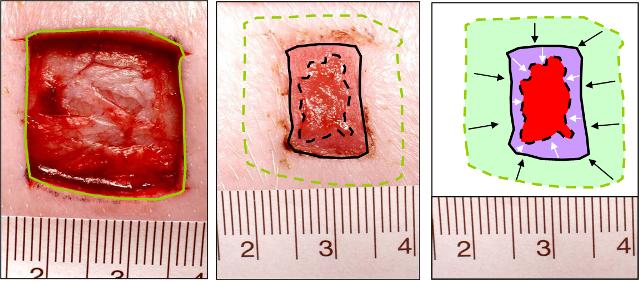
Wound Closure differentiated into: Wound Contraction (green area) & Wound Re-epithelialisation (purple area). Red area = open wound.
|
|
B, Wound Healing Progression Wound healing progression can be determined in a number of ways. We evaluate the impact of agents on wound healing progression in terms of:- 1. Wound Colour Profile 2. Cellular Profile of Wound Tissue
|
|
A. Rate of Wound Closure The impact of agents on wound closure is assessed using excisional wound models. Wound closure, which is measured by computer-assisted planimetry from calibrated images taken at intervals during the study, can be differentiated into its components:- Wound Re-epithelialisation & Wound Contraction (see below)
|
|
Neutrophils (Chloroacetate Esterase staining) |
|
Macrophages (ED-1 immuno-staining) |
|
Angiogenesis (anti-CD31 immuno-staining) immuno-staining) |
|
C. New Tissue Formation [information to be inserted June 2009) |
|
2. Cellular Profile of Wound Tissue Under normal “uncompromised” circumstances skin wounds are “seen” to progress through the stages of inflammation, new tissue formation and new tissue remodelling - which results in the formation of scar tissue (a virtually avascular & acellular tissue functionally and aesthetically inferior to normal skin). The cellular profile of wound tissue changes with time after injury. Initially, inflammatory cells that are responsible for clearing wound site debris (such as neutrophils and monocyte-macrophages) predominate; later as the wound heals (matures) proliferative cells which are responsible for the generation of replacement tissues (i.e. fibroblasts & endothelial cells) predominate. Consequently, the cellular profile of a wound can be used to describe its maturity. Wound tissues with a relatively high proportion of inflammatory cells are considered less mature (less advanced) than similar tissues containing relatively few inflammatory cells. The relative contribution of inflammatory cells (neutrophils & monocyte-macrophages) and proliferative cells (fibroblasts & endothelial cells) to wound cellularity—at a given point in time after injury and treatment — allows the impact of an investigational agent on wound progression to be determined. Cell specific histological staining techniques are used to label and measure the involvement of specific cellular lineages (see below).
|
|
1. Wound Colour Profile (CIELAB Wound Colour Analysis] [information to be inserted June 2009) |
|
Assessment of Impact of Investigational Agents/Therapies on Wound Healing The impact of investigational agents / therapies on wound healing can be assessed using a variety of measurement parameters and endpoints.
The following, clinically relevant, assessments are routinely undertaken:- A. Rate of Wound Closure B. Wound Healing Progression (incomplete] C. New Tissue Formation [incomplete] D. Tensile Strength [incomplete] E. Scarring
|
|
see text below for details |
|
D. Tensile Strength [information to be inserted June 2009) |
|
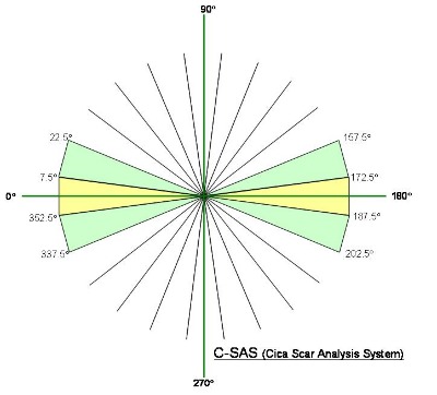
Scar Tissue Analysis using the Cica Scar Analysis System (C-SAS) C-SAS generates data describing the directionality of collagen bundles within scar dermal tissue. The output generated by C-SAS describes the directionality of collagenous structures in twelve 15° segments (see graphic).
|